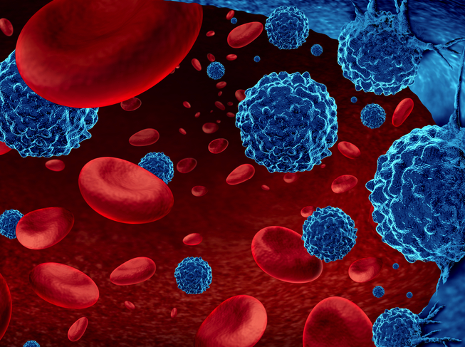
Glioblastoma is a very aggressive form of brain cancer that will often infiltrate surrounding brain tissue, making treatment extremely difficult. Now, in a new study conducted by scientists at Sanford-Burnham Medical Research Institute and the Salk Institute for Biological Studies, both in the United States, a method that combines a tumor-homing peptide, a cell-killing peptide and a nanoparticle that both enhances tumor death and allows researchers to image the tumors, has been developed. The new nanosystem, when used to treat mice with glioblastoma, eradicated most tumors in one model, and significantly delayed tumor development in another. Erkki Ruoslahti, M.D., Ph.D., the senior author of the study and distinguished professor in both Sanford-Burnham’s NCI-designated Cancer Center and the Center for Nanomedicine, a Sanford-Burnham collaboration with the University of California, Santa Barbara, said, “This is a unique nanosystem for two reasons. First, linking the cell-killing peptide to nanoparticles made it possible for us to deliver it specifically to tumors, virtually eliminating the killer peptide’s toxicity to normal tissues. Second, ordinarily researchers and clinicians are happy if they are able to deliver more drugs to a tumor than to normal tissues. We not only accomplished that, but were able to design our nanoparticles to deliver the killer peptide right where it acts—the mitochondria, the cell’s energy-generating center.” Treatment with the nanosystem cured all but one of the ten mice with the glioblastoma. Additionally, because the nanoparticles are made of iron oxide, they are visible by MRI, so they could aid in diagnosing the disease. The team’s findings were published in the Proceedings of the National Academy of Sciences of the USA.
http://www.sanfordburnham.org/news_and_events/news_archive/2011/october_4_2011_erkki.aspx